Efficacy of Stilbene Derivatives in Sensitizing Breast Cancer Cells to Ionizing Radiation
Keywords
Abstract
Background/Aims:
One of the treatments for breast cancer is surgical resection of the tumour and prevention of recurrence with postoperative radiotherapy. Unfortunately, radiotherapy is not always effective enough due to the low sensitivity of cancer cells to ionising radiation. This study aimed to evaluate the radiosensitising properties of resveratrol, piceatannol and polydatin on breast cancer cells, which differ in the presence of hormonal receptors on their surface.Methods:
The experimental part was carried out on triple-negative breast cancer cells (HCC38) and hormone-dependent cells (MCF7). The study assessed the level of cell death, changes in the expression of genes (BAX, BCL-2) and proteins related to the apoptosis process (CASPASE 3, 8 and P53), changes in the expression of antioxidant enzymes (CATALASE, SOD, GPx1/2) and NRF-2. Additionally, the expression level of RAD51 protein and histone H2AX, which are involved in DNA repair processes, was assessed. Statistical significance was evaluated by a two-way analysis of variance (ANOVA) followed by Tukey’s post hoc test (p < 0.05).Results:
Ionising radiation in combination with resveratrol or piceatannol activates the apoptosis process by internal and external pathways. Greater sensitivity of MCF7 cells compared to HCC38 cells to ionising radiation in combination with resveratrol is associated with a weaker antioxidant response of cells and reduced intensity of DNA damage repair. DNA repair induced by ionising radiation occurs more effectively in HCC38 cells than in MCF7 cells.Conclusion:
Resveratrol has the highest radiosensitising potential among the tested stilbene for cells of both lines. The effectiveness of ionizing radiation in combination with resveratrol (to a lesser extent with piceatannol) is more significant in MCF7 than in HCC38 cells.Introduction
Cancer is the second leading cause of death in the world. According to the oncology report of the World Health Organization, the greatest threats to human life and health are breast and lung cancer, and the number of cases of colon, prostate, stomach and liver cancer is also increasing. Both in the prevention and treatment of various cancers, compounds of natural origin play a vital role [1–3]. It is estimated that 60 to 80% of drugs used to treat cancer are derived directly or indirectly from naturally occurring species [3, 4].
Currently, several studies are carried out on compounds of natural origin that would sensitise cancer cells to ionising radiation [5, 6]. Compounds from the polyphenols group are of particular interest and are the subject of much research. Several representatives of this group have anticancer activity. The advantage of these compounds over synthesised synthetic agents is their widespread availability, relatively low acquisition costs and well-characterised safety profile.
The most well-known compound from the group of polyphenols is the stilbene derivative - resveratrol (3, 4,5-trans-trihydroxystilbene, abbreviated R) (Fig. 1). It was first discovered by Takaoka in Veratrum grandiflorum in 1940 [7]. It is characterised by solid antioxidant, neuroprotective, anti-inflammatory, and anticancer properties [8–11]. Unfortunately, the main problem with using resveratrol in clinical treatment is its poor bioavailability. The gastrointestinal tract strongly absorbs, rapidly metabolises and excretes it in the urine [12]. To solve this problem, effective carriers are being sought that would allow resveratrol to be delivered to cells in a higher concentration. Research is also conducted on resveratrol analogues, which, thanks to minor changes in the structure, are characterised by much greater bioavailability [13].
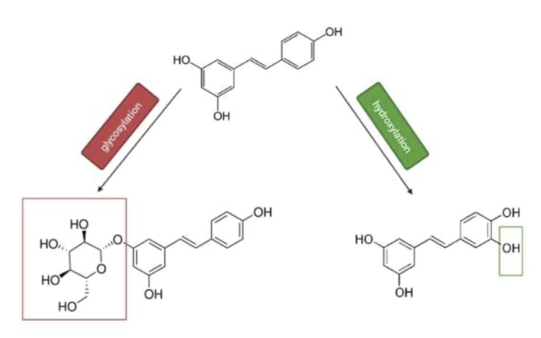
Fig. 1: Chemical structure of resveratrol (on the top), polydatin (on the left) and piceatannol (on the right).
One such compound is one of the resveratrol metabolites - piceatannol (3-hydroxyresveratol, abbreviated as ROH). Piceatannol differs from resveratrol by adding a hydroxyl group in the 3’ position. Previous studies conducted with piceatannol showed it is more stable in the metabolic process than resveratrol [14]. In addition, studies using ROH have demonstrated that it exhibits several bioactivities, such as anti-inflammatory, antioxidant and anticancer effects [15]. Another compound from the stilbene group, with better pharmacokinetic properties than resveratrol, is the precursor of resveratrol - polydatin, also called piceid (glucoside of resveratrol in which the glucoside group bound to the C-3 position substitutes a hydroxyl group, abbreviated as RG). Previous studies have shown that polydatin can retain the biological properties of resveratrol but is less susceptible to enzymatic oxidation. In addition, polydatin is much more soluble in water than resveratrol [16, 17].
Breast cancer is one of the most common cancers in women and has the highest mortality rate. Despite many years of research, the incidence of breast cancer continues to rise in some countries. Every 20th woman in the world and every 8th in developed countries has breast cancer [18]. In 2020, the number of cases amounted to 2.3 million, of which 685 thousand patients died. Unfortunately, the prognosis is not very promising; in 2070, the number of breast cancer cases is expected to increase to 4.4 million [19]. In the case of breast cancer, recurrences are also a problem, as approximately 30% of patients diagnosed with early-stage cancer have recurrence [20]. Breast cancer can be divided into three types. This division concerns the molecular markers of progesterone (PR) or estrogen (ER) receptors (their presence or absence) and the presence or absence of human epidermal growth factor receptor 2 (ERBB2; formerly HER2). The most common subtype of BC is the presence of hormone receptors and ERBB2 negative, which occurs in approximately 70% of patients. The hormone-positive and ERBB2-positive subtype accounts for about 15-20% of all cases. In contrast, the triple-negative subtype (TNBC), i.e. one in which none of the three molecular markers is present, accounts for approximately 15% of the cases [21]. Triple-negative breast cancer cells are much more invasive and aggressive than others [22].
Most patients with breast cancer, as much as 90% of cases, do not have metastases at the time of diagnosis [21]. In such patients, treatment is mainly based on surgical resection of the tumour and prevention of recurrence with postoperative radiotherapy (RT), chemotherapy or hormone therapy, depending on the tumour subtype [23]. However, relapses are more frequent in patients with the triple-negative breast cancer subtype than in patients with the other two BC subtypes [24, 25]. Treatment of patients with metastatic breast cancer is closely related to the cancer subtype and aims to prolong life and improve its comfort by alleviating the symptoms of the disease. The worst prognosis for breast cancer is the triple-negative subtype, with a median survival of about one year, compared to the other two subtypes, where the median survival is five years [21].
As mentioned above, radiotherapy is also used to treat breast cancer. It is used as a permanent element of sparing treatment after surgical tumour removal. After mastectomy, postoperative radiotherapy reduces the number of local recurrences and increases the likelihood of long-term survival. In cases where the advancement of the cancer or the patient’s general condition does not allow for surgical treatment, radiotherapy is the only method of treating local breast cancer. Unfortunately, RT is not always effective enough, which may be due to the low sensitivity of cancer cells to ionising radiation (IR) [26].
Therefore, we decided to check whether resveratrol and its two derivatives (piceatannol and polydatin) can enhance the effect of ionising radiation on triple-negative breast cancer cells (HCC38; ER- PR-, HER2-) and hormones dependent cells (MCF7; ER+, PR+, HER2-). The conducted research also allowed us to indicate which of the tested com-pounds enhances the effect of ionising radiation to the greatest extent.
Materials and Methods
Cell Line
The experiments were conducted on the human breast carcinoma cell line MCF7 (hormone-dependent cell) and HCC38 (triple-negative breast cancer cell line). Both lines were obtained from ATCC (Manassas, VA, USA). MCF7 were grown in Dulbecco’s modified Eagle’s medium (DMEM) supplemented with 10% fetal bovine serum and antibiotics (10U/mL penicillin and 50μg/mL streptomycin). HCC38 were cultivated in Roswell Park Memorial Institute medium (RPMI-1640) supplemented with 10% fetal bovine serum and antibiotics (10 U/mL penicillin and 50 μg/mL streptomycin). Cells were grown in standard conditions (37°C, 5% CO2). After reaching 80-90% confluence, cells were carefully removed with trypsin/EDTA and washed with fresh phosphate-buffered saline (PBS). Cell viability was determined using the trypan blue assay.
Chemicals and Reagents
Cell culture media, antibiotics (penicillin/streptomycin), and fetal bovine serum were purchased from Sigma-Aldrich. Reagents for detecting apoptosis were purchased from Molecular Probes. The reagent for the RNA EXTRACTME isolation kit was obtained from BLIRT. RevertAid First Strand kit for cDNA synthesis was purchased from Applied Thermo Scientific. 5x HOT FIREPol® EvaGreen® qPCR Supermix comes from Solis Biodyne. Specific antibodies for BCL-2, CASPASE 3, 8, SOD1 and SOD2, GPX1/2, CATALASE, P53, NRF2, RAD51, H2A.X and β-ACTIN and secondary antibodies were obtained from Santa Cruz Biotechnology. Oligonucleotide, Bax, Bcl-2 and hypoxanthine phosphoribosyl transferase (HPRT) were obtained from the Genomed firm.
Three stilbene derivatives were used in the experiment: resveratrol (3, 4′,5-trans-trihydroxystilbene, R), (Sigma-Aldrich (St. Louis, MO, USA)), piceatannol (3, 3′,4, 5′-trans-trihydroxystilbene, ROH) (Cayman Chemical (Ann Arbor, MI, USA)) and polydatin (3, 4′,5-trans-trihydroxystilbene-3-O-β-mono-d-glucoside, RG) - the glucoside form of resveratrol (Sigma-Aldrich (St. Louis, MO, USA)). All stilbene compounds were dissolved in ethyl alcohol to the specified initial concentration. The concentration was determined spectrophotometrically at a wavelength of 304 nm in the case of resveratrol and polydatin and 326 nm in the case of piceatannol. The concentrations of the compounds were calculated based on molar absorption coefficients: for resveratrol and polydatin, this value was 30, 335 M-1cm-1 [27], and for piceatannol 33, 100 M-1cm-1 [28]. Depending on the type of experiment, the compounds were diluted to a specific concentration and added to the cell medium.
Detection of Apoptosis and Necrosis by Flow Cytometry
Apoptotic, necrotic, and living cells were quantified by double staining with the Annexin V Apoptosis Detection Kit II, employing FITC-labelled Annexin V. Annexin V binds to cells that expose phosphatidylserine at their surface, a feature of cells that are undergoing apoptosis.
Cells were seeded on sterile dishes (Ø 40 mm) in 1, 000, 000 for MCF7 cells and 500, 000 for HCC38 cells, then incubated for 24 hours. After this time, the cells were preincubated with resveratrol or its derivatives (piceatannol or polydatin) at a concentration of 25 µM (MCF7 line) or 50 µM (HCC38 line) for 3 hours at 37°C. Then, they were exposed to ionising radiation at a dose of 6 Gy. Cells were incubated for 24 h and then trypsinized, washed with DPBS, and suspended in 1× binding buffer to which 5 μl of FITC annexin and 5 μl of propidium iodide were added. The cells were incubated for 15 minutes in the dark, and then fluorescence was measured within an hour. All fluorescence measurements were performed on a Becton Dickinson LSR II cytometer. Cells emitting weak green fluorescence (FITC annexin) and weak red fluorescence (propidium iodide) were counted as apoptotic.
Gene Expression Analysis by Real-Time PCR
Total RNA was extracted using the Extractme Total RNA kit according to the manufacturer’s instructions. The concentration of isolated RNA is determined using a NanoDrop spectrophotometer. All RNA samples obtained were stored at -80°C for further analysis. Approximately 1 μg of extracted RNA was reverse transcribed into cDNA using a reverse transcription kit (RevertAid first-strand cDNA synthesis kit) according to the protocol established by the manufacturer. The cDNA sample was stored at −20°C until use. The BCL-2 and BAX genes were expressed using an Eva Green probe (Solis Biodyne, Tartu, Estonia). The HPRT gene was a reference gene. Sequences of PCR primers used for RT-PCR with expected product size are listed in Table 1. Polymerase chain reaction during the reaction using the Eco 48 Real-time PCR thermal cycler. The preparation consisted of: 2 µl EvaGreen Supermix, 1 µl cDNA and 0.2 µl of each set of primers, 6.6 µl H2O. All were amplified three times. The final profile was 12 s at 95°C and 40 cycles of 95°C for 15 s and 60°C for 20 s.
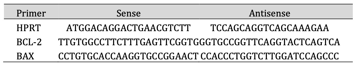
Table 1: Primer sequenes used for real-time quantitative reverse transcriptase polymerase chain reaction (qRT-PCR)
Western Blot and Densitometric Analysis
Cell preparations were lysed in RIPA buffer (Sigma–Aldrich (St. Louis, MO, USA)) supplemented with AEBSF protease inhibitor cocktail, aprotinin, bestatin, E-64, leupeptin, and pepstatin A (Thermo Scientific 100 × Halt protease inhibitor cocktail). Then, they were incubated on ice for 30 minutes. After incubation on ice, the samples were centrifuged at 5000 rpm for 5 minutes at 4°C. Protein concentration in cell preparations was determined using the Lowry method. The separation of protein in cell lysates was performed using 12.5% SDS-PAGE. Proteins separated in a polyacrylamide gel using the Towbin method were transferred electrophoretically to an Immobilon-P membrane (pore diameter 0.45 µm). Proteins immobilized on the Immobilon-P membrane were subjected to immunodetection. To detect proteins on immunoblots, specific primary antibodies were used: an-ti-BCL-2 (sc-7382), anti-CASPASE-3 (sc-7272), anti-CASPASE-8 (sc-56070), anti-GPx1/2 (sc-133160), anti-SOD1 (sc-17767), anti-SOD2 (sc-133134), anti-CATALASE (sc-271803), anti-P53 (sc-393031), anti-NEF2 (sc-365949), anti-RAD51 (sc-56212), anti-H2A.X (sc-517336). M-IgGκ BP (sc-516102) were used as secondary antibodies. For loading, the control detection of β-ACTIN was performed by an anti-β-ACTIN antibody (sc-477778). All antibodies were purchased from Santa Cruz Biotechnology, Santa Cruz, CA, USA. The resulting complex (antigen-antibody) was identified using the chemiluminescence method using the SuperSignal™ West Pico PLUS Chemiluminescent Substrate kit (Thermofisher ScientificTM, Lithuania). The analysis was performed using a high-performance Western blot system - Azure Imaging Systems (Syngen, Wrocław, Poland). The results obtained at the level of the tested proteins were subjected to densitometric analysis in the Image J program. The results were presented as average integrated optical density (IOD) values. The optical density values obtained for the bands corresponding to individual proteins were divided by the optical density value obtained for the β-ACTIN band.
Statistical Analysis
All results are presented as the mean ± SD of three independent repetitions. Treatments were compared by a two-way analysis of variance (ANOVA) followed by Tukey’s post hoc test (p < 0.05).
Results
The concentrations of the compounds that were tested in this study were determined based on the level of apoptosis assessed by the cytofluorimetric method of Annexin V-FITC/PI staining. The concentration that was selected the one at which resveratrol combined with ionising radiation (6Gy) induced a comparable level of apoptosis (approximately 33%) in both cell lines. For MCF7 cells, the concentration was 25 µM, and for the HCC38 cell line, it was 50 µM.
Stilbene derivatives alone and in combination with radiation promote the activation of caspases and induce apoptosis of breast cancer cells
The level of apoptosis in MCF7 and HCC38 cells treated with stilbene derivatives and combined with ionising radiation is shown in Fig. 2.
The results showed that MCF7 cells are slightly more sensitive to radiation than HCC38 cells (apoptosis levels are approximately 20% and 10% for MCF7 and HCC38, respectively). The highest level of apoptosis (approximately 33%) was observed in cells of both lines irradiated with a dose of 6Gy, which were previously incubated with resveratrol (25 µM for MCF7 cells and 50 µM for the HCC38 cell line). For the HCC38 line, a similar level of apoptosis was obtained in cells preincubated with piceatannol. The next task was to determine the molecular mechanism by which apoptosis is activated.
We examined protein expression levels involved in apoptosis: BCL-2, P53, CASPASE 3 and 8 (Figures 3 and 4). We also attempted to evaluate BAX protein expression, but we obtained no results in any tested cell lines (HCC38 and MCF7). We conclude that the purchased antibodies were of poor quality. Therefore, we additionally determined the expression level of the BAX and BCL-2 gene. The BAX to BCL-2 expression ratio determines cells’ life or death in response to an apoptotic stimulus; an increased BAX /BCL-2 ratio decreases the cellular resistance to apoptotic stimuli, leading to increased cell death [29]. Discussion of the BAX/BCL-2 ratio is a better indicator of further cell fate than analysing the level of only one of these proteins.
In MCF7 cells, we observed an increase in the BAX /BCL-2 ratio in all systems tested. The most significant BAX/BCL-2 ratio increase was observed when the cell was treated with resveratrol combined with IR (Fig. 3B). In the same system, we also observed the highest level of apoptosis.
The level of the anti-apoptotic protein BCL-2 slightly increases in all systems tested except for cells treated with polydatin and then irradiated compared to control cells (cells not treated with any of the tested factors). It should be emphasised, however, that in systems where cells were treated with the tested compounds in combination with radiation, the protein level decreased compared to cells irradiated solely (Fig. 3C).
In none of the tested systems, MCF7 cells had an increased level of P53 protein compared to the control. This means that apoptosis is activated independently of the P53 protein in MCF7 cells (Fig. 3D).
The studies performed showed significant increases in the level of CASPASE 8 in MCF7 cells treated with radiation alone, with the compounds alone, and with resveratrol or piceatannol in combination with IR (Fig. 3E).
The study also attempted to assess the level of CASPASE 3, but as expected, this protein was not detected in MCF7.
In HCC38 cells, the BAX/BCL-2 gene expression ratio increases in all systems but to the greatest extent in cells treated with resveratrol or piceatannol and combined with ionising radiation (Fig. 4B).
BCL-2 protein levels, assessed by Western Blot, increased slightly in cells treated with piceatannol or polydatin and polydatin combined with IR. In other systems, its level is close to control values (cells not treated with any tested factors) (Fig. 4C).
P53 protein levels increase in almost all systems tested except polydatin and IR-treated cells (Figure D). Moreover, in the case of this protein, we observe a trend of changes similar to those observed in the BAX/BCL-2 expression ratio (Fig. 4B).
The most significant increase in CASPASE 3 levels was observed in HCC38 cells preincubated with resveratrol or piceatannol in combination with ionising radiation (Fig. 4E). The highest levels of CASPASE 8 were observed in cells exposed only to irradiation and incubated with piceatannol combined with IR (Fig. 4F). In the case of polydatin used in combination with IR, a significant decrease in CASPASE 3 and 8 levels was observed.

Fig. 2: The level of apoptosis was assessed based on the cytofluorimetric method using Annexin V-FITC/PI staining. (A) the MCF7 cells were treated with 25 μM of resveratrol (R), piceatannol (ROH), or polydatin (RG) alone and in combination with IR (dose of 6 Gy). Own data, adapted from [26]. (B) the HCC38 cells were treated with 50 μM of resveratrol (R), piceatannol (ROH), and polydatin (RG) alone and in combination with IR (dose of 6 Gy). Data are shown as the mean ± SD of three independent repetitions. The mean difference (*) was compared with the control and (#) with IR.
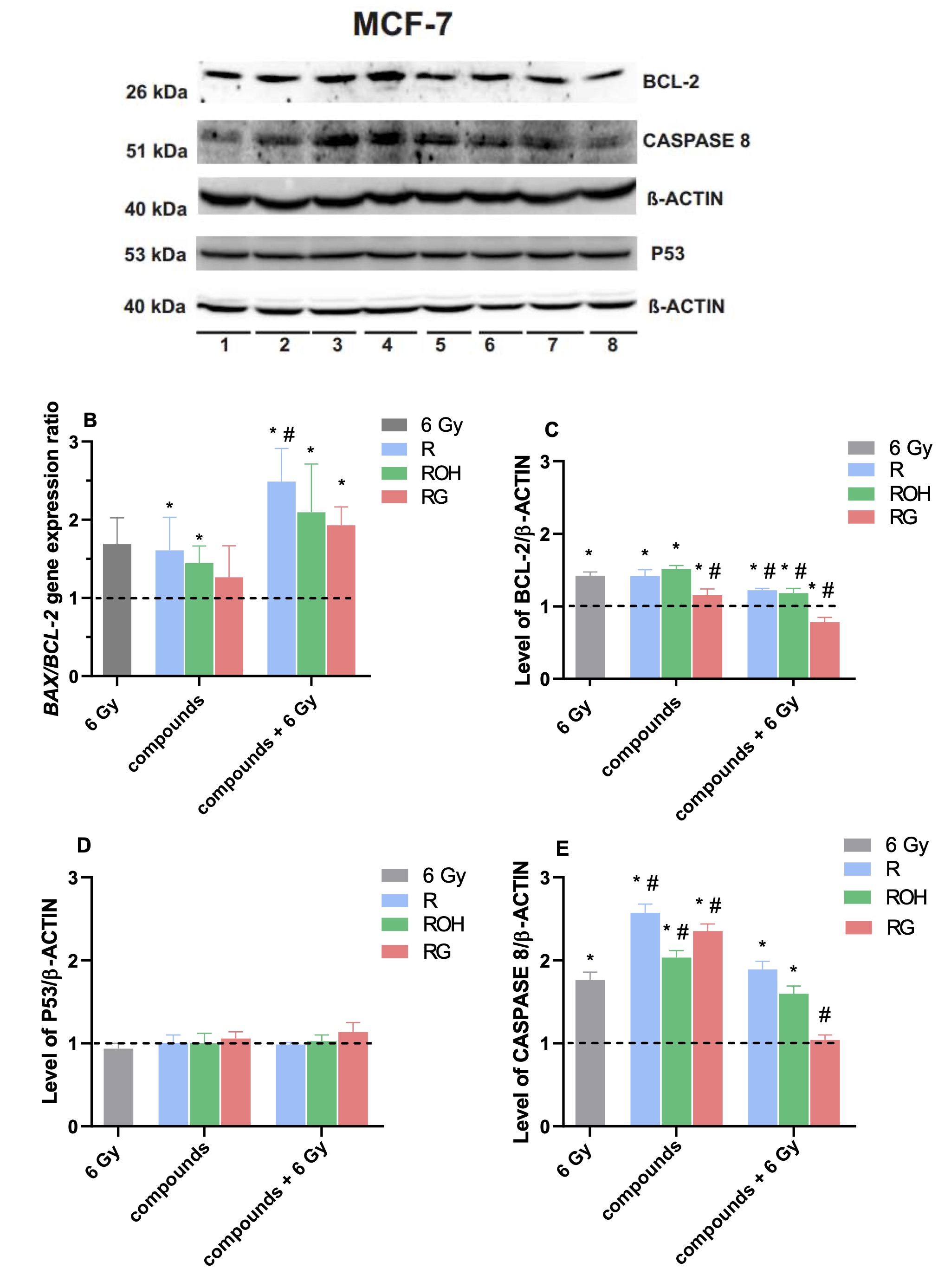
Fig. 3: Expression level of selected genes and proteins involved in the apoptosis process in MCF7 cells: (A) representative image of western blotting results obtained in this study, lines: (1) – control cells, (2) – cells irradiated with 6 Gy, next cells treated with: (3) – resveratrol, (4) – piceatannol, (5) – polydatin, and (7) – resveratrol and 6 Gy, (8) – piceatannol and 6Gy, (9) – polydatin and 6 Gy. (B) the BAX/BCL2 gene expression level ratio assessed based on the real-time PCR method—own data, adapted from [26]. Relative level of proteins identification by Western blot method: (C) BCL-2 protein, (D) P53 protein, and (E) CASPASE 8 in MCF7 cells treated with 25 μM of resveratrol (R), piceatannol (ROH), and polydatin (RG) alone and in combination with IR (dose of 6 Gy). The results obtained in each kind of sample were compared to controls, i.e., cells not treated with any of the tested factors. The mean difference (*) was compared with the control and (#) with IR.
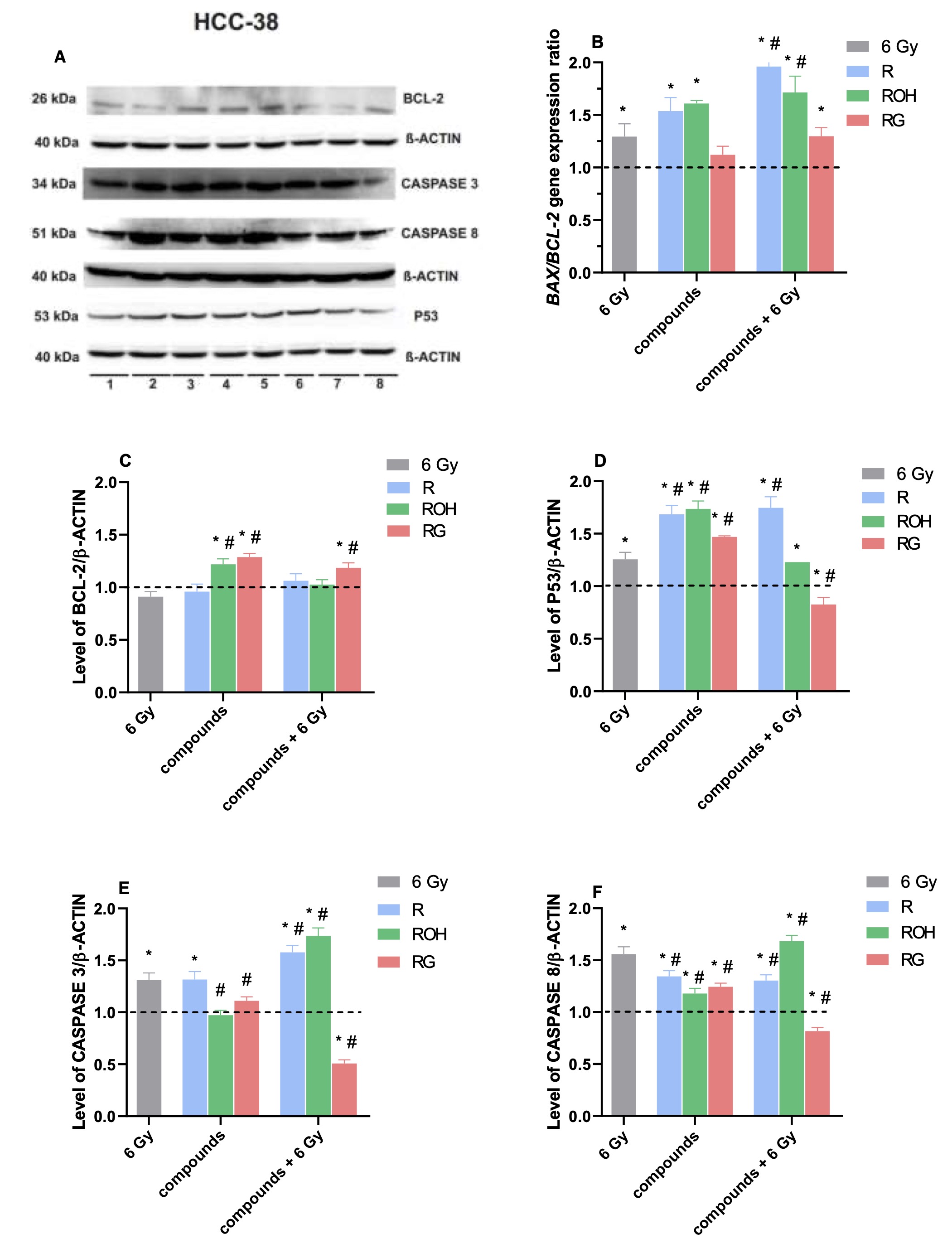
Fig. 4: Expression level of selected genes and proteins involved in the apoptosis process in HCC38 cells: (A) representative image of western blotting results obtained in this study, lines: (1) – control cells, (2) – cells irradiated with 6 Gy, following cells treated with: (3) – resveratrol, (4) – piceatannol, (5) – polydatin, and (7) – resveratrol and 6 Gy, (8) – piceatannol and 6Gy, (9) – polydatin and 6 Gy. (B) BAX/BCL2 gene expression level ratio assessed based on the real-time PCR method. Relative level of protein identification by Western blot method: (C) BCL-2 protein, (D) P53 protein, (E) CASPASE 3, and (F) CASPASE 8 in HCC38 cells treated with 50 μM of resveratrol (R), piceatannol (ROH), and polydatin (RG) alone and in combination with IR (dose of 6 Gy). The results obtained in each kind of sample were compared to controls, i.e., cells not treated with any of the tested factors. The mean difference (*) was compared with the control and (#) with IR.
Stilbene derivatives alone and in combination with radiation affect the level of antioxidant enzymes
The primary assumption of radiotherapy is to induce the death of cancer cells by generating reactive oxygen species (ROS). Therefore, reducing the antioxidant potential of cancer cells should enhance the effectiveness of radiotherapy. The study examined how stilbene compounds used alone or combined with ionising radiation affect the expression level of antioxidant enzymes: catalase, cytoplasmic (SOD1) and mitochondrial (SOD2) superoxide dismutases and glutathione peroxidases (GPX1/2). The level of NRF2, an essential protein in the cell’s antioxidant defence response mechanism, was also assessed (Figures 5 and 6).
Superoxide dismutases
In MCF7, a statistically significant, approximately two-fold increase in SOD1 level was observed in all tested systems (Fig. 5B). However, there were no changes in the SOD2 level compared to the control (Fig. 5C). The response of HCC38 cells to changes in SOD1 and SOD2 levels in our studies was generally opposite to the response observed for MCF7 cells (Figures 6 B and C). The status of SOD1 did not change significantly or decrease by about 20% and 40% for cells treated with resveratrol alone and polydatin combined with IR, respectively. In one case, cells treated with piceatannol and IR showed an increase in SOD1 level (about 40% vs. control). In the SOD2 level, a significant increase was noted in cells treated with resveratrol or piceatannol, also in combination with ionising radiation. The effect of resveratrol was approximately 1.5 times stronger than that of piceatannol. Radiation, whether used alone or combined with stilbenes, does not affect changes in SOD2 expression.
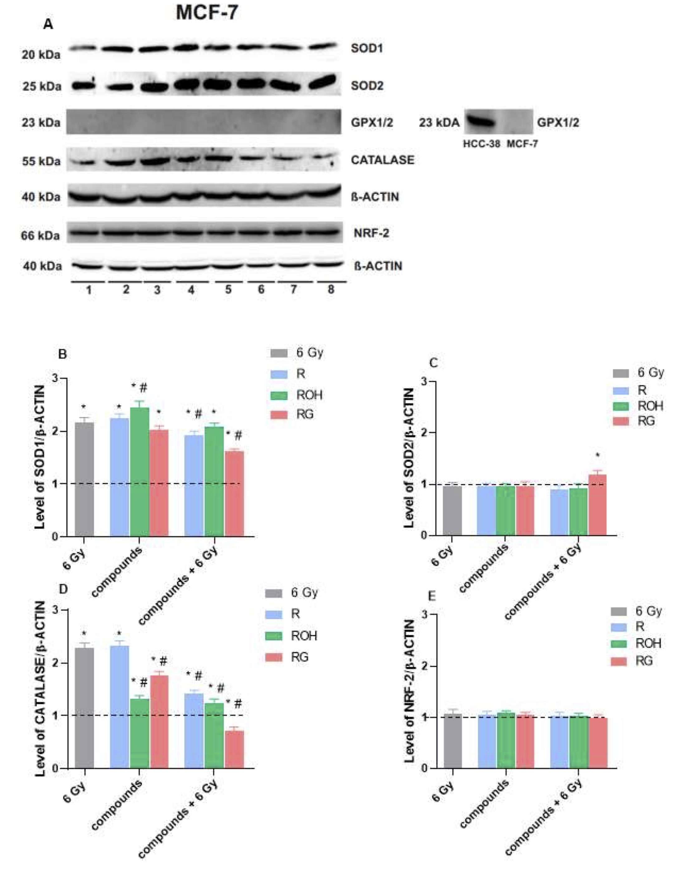
Fig. 5: Immunoblot assay to detect the expression level of antioxidant enzymes in MCF7 cells: (A) representative image of western blotting results, lines: lines: (1) – control cells, (2) – cells irradiated with 6 Gy, next cells treated with: (3) – resveratrol, (4) – piceatannol, (5) – polydatin, and (7) – resveratrol + 6 Gy, (8) – piceatannol + 6Gy, (9) – polydatin + 6 Gy. (B) level of SOD1, (C) level of SOD2, (D) level of catalase and (E) level of NRF-2 factor in cells treated with 25 μM of resveratrol (R), piceatannol (ROH), and polydatin (RG) alone and in combination with IR (dose of 6 Gy). The results obtained in each kind of sample were compared to controls, i.e., cells not treated with any of the tested factors. The mean difference (*) was compared with the control and (#) with IR.
Catalase
In both cell lines, an increase in catalase levels was observed in all systems tested except one in which MCF7 cells were treated with polydatine and IR (Figures 5D and 6D). The main difference between MCF7 and HCC38 concerns the response of cells preincubated with resveratrol or piceatannol in combination with IR compared to cells subjected to irradiation only. In the case of MCF7, the level of catalase in cells preincubated with compounds in combination with IR is much lower than in cells only irradiated. In HCC38, we observe the opposite tendency, i.e. in cells treated with resveratrol or piceatannol and then irradiated, the enzyme level is approximately 2 times higher compared to cells only irradiated.
Glutathione peroxidases
The last enzyme analysed was the glutathione peroxidase level, isoenzyme 1 and 2. We did not detect these proteins in MCF7 cells (Fig. 5, on the right side of drawing A), probably due to their absence or low level in cells of this line, which other authors previously noticed. Kulak et al. observed low levels of GPx1 mRNA but did not demonstrate the presence of the protein by Western Blot [30]. In turn, Chu et al. reported GPx2 activity but showed deficient cell protein levels [31]. In HCC38 cells, the greatest changes, consisting of a significant reduction of the enzyme level, were observed in cells pre-incubated with the tested stilbenes and then subjected to irradiation. The most significant reduction of enzyme levels was observed in cells treated with resveratrol in combination with radiation (Fig. 6E).
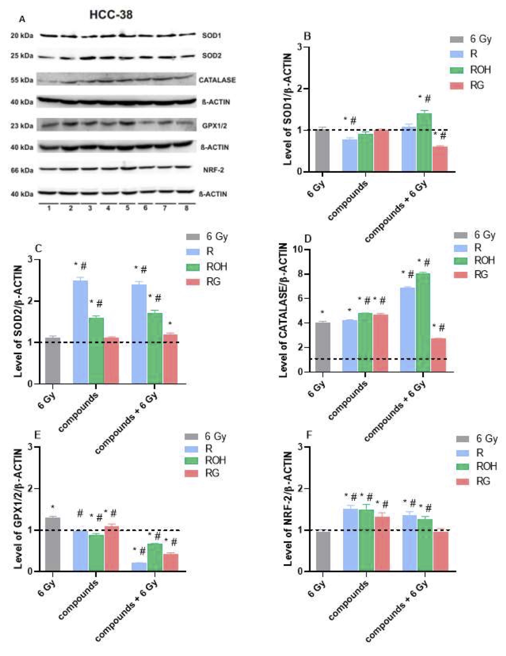
Fig. 6: Immunoblot assay to detect the expression level of antioxidant enzymes in HCC38 cells: (A) representative image of western blotting results, lines: lines: (1) – control cells, (2) – cells irradiated with 6 Gy, next cells treated with: (3) – resveratrol, (4) – piceatannol, (5) – polydatin, and (7) – resveratrol + 6 Gy, (8) – piceatannol + 6Gy, (9) – polydatin + 6 Gy, (B) level of SOD1, (C) level of SOD2, (D) level of catalase, (E) level of GPX1/2 and (F) level of NRF-2 factor in cells treated with 50 μM of resveratrol (R), piceatannol (ROH), and polydatin (RG) alone and in combination with IR (dose of 6 Gy). The results obtained in each kind of sample were compared to controls, i.e., cells not treated with any of the tested factors. The mean difference (*) was compared with the control and (#) with IR.
Nuclear factor-erythroid-2–related factor 2 (NRF-2)
In MCF7 cells, no changes in NRF2 levels were observed in any of the tested systems compared to the control (Fig. 5E). In HCC38 cells, ionising radiation did not affect the expression level of NRF2, while stilbene derivatives alone increased expression by approximately 1.5 times on average (Fig. 6F). Using resveratrol or piceatannol combined with ionising radiation increased the level of the NRF2 factor but slightly less than the effects of the compounds alone.
Activation of proteins responsible for repairing damaged DNA is more effective in HCC38 than in MCF7 cells
The radiosensitivity of cancer cells largely depends on the effectiveness of repairing damaged DNA. Therefore, the study determined the expression levels of two proteins involved in DNA repair processes - RAD51 protein and histone H2AX (Fig. 7). RAD51 is a recombinase that mediates the repair of double-stranded DNA breaks by homologous recombination (HR) [32]. In turn, the level of H2AX expression is a parameter indicating the intensity of induction of the DNA repair process, tumour aggressiveness, and the prognosis of breast cancer patients. According to the authors Katsuta et al. (2022), the expression level of H2AX is as good a parameter as the most frequently analysed level of phosphorylation of this histone (γ-H2AX) [33].
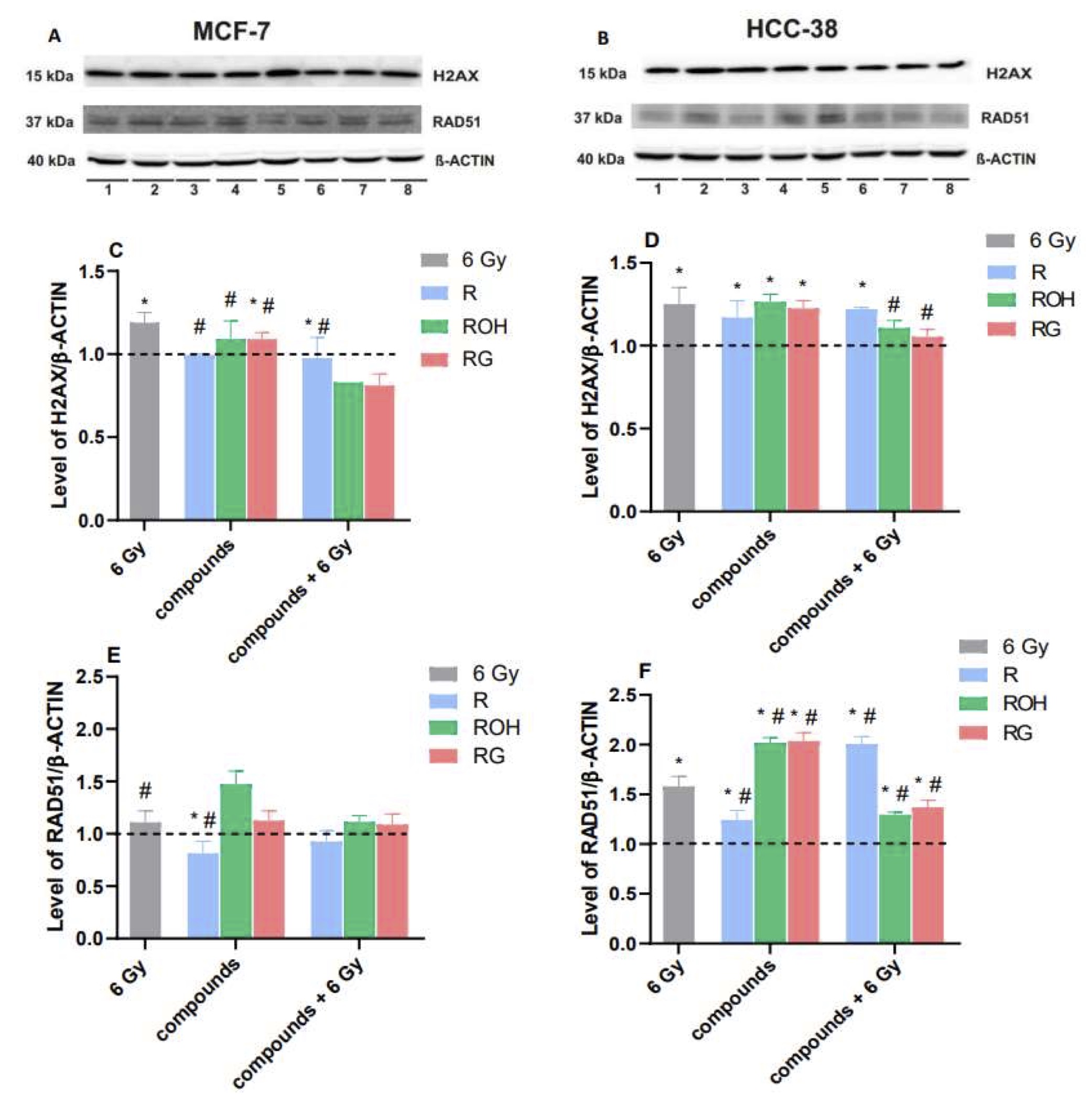
Fig. 7: Immunoblot test detecting the expression level of two proteins involved in DNA repair processes - RAD51 protein and histone H2AX in cells of both lines: (A and B) representative image of Western blot results for MCF7 and HCC38 cells, respectively; the lines on both blots represent: (1) – control cells, (2) – cells irradiated with 6 Gy, and cells treated with: (3) – resveratrol, (4) – piceatannol, (5) – polydatin, (6) – resveratrol and 6 Gy, (7) – piceatannol and 6Gy, (8) – polydatin + 6 Gy. (C and D) H2AX level in MCF7 and HCC38 cells, respectively, (E and F) RAD51 level in MCF7 and HCC38 cells, respectively. Cells were treated with stilbene derivatives at a concentration of 25 µM for the MCF7 line and 50 µM for the HCC38 line. The results obtained in each kind of sample were compared to controls, i.e., cells not treated with any of the tested factors. The mean difference (*) was compared with the control and (#) with IR.
The results indicate that the tested stilbene derivatives alone and in combination with IR did not enhance, and in some cases even reduce, the RAD51 and H2AX protein expression levels in MCF7 cells. The only instances in which we observed an increase in the level of RAD51 protein, compared to the control, were in cells treated with piceatannol alone. HCC38 cells showed an increase in RAD51 levels in all systems. In cells treated with resveratrol alone, the increase in protein levels was relatively small and amounted to approximately 20%. They significantly increased by about 80% when cells were additionally treated with radiation. Opposite trends were observed in cells treated with piceatannol and polydatin, i.e. the compounds alone significantly increased protein expression, while in combination with radiation, the RAD51 level significantly decreased. In the case of changes in the expression level of H2AX, the nature of the observed changes was similar to that of the RAD51 protein, but the expression level of H2AX was much lower.
Discussion
The study compared the radiosensitising properties of resveratrol and its two derivatives: piceatannol and polydatin. The choice of monohydroxylated and glucosylated derivatives of resveratrol was dictated by their greater bioavailability than resveratrol [34, 35]. Moreover, it resulted from the lack of literature data regarding their use, in combination with ionising radiation, on cancer cells.
The study does not present the results of metabolic viability obtained using methods based on tetrazolium salt reduction (MTT or MTS test). Our observations and other authors indicate that ionising radiation affects the biogenesis and hyperactivation of mitochondria. Hence, metabolic viability studies in experiments with ionising radiation do not give an accurate picture of cell viability compared to the actual number of cells [36].
The concentrations of compounds used in the studies for a given cell line were selected based on the determination of the level of apoptosis in cells treated with resveratrol and ionising radiation. This concentration caused a comparable level of cell death for both cell lines, i.e. 25 µM for MCF7 cells and 50 µM for HCC38 cells. According to literature data, these concentrations are not toxic or have low toxicity for normal cells [37–40]. In many types of cancer cells, resveratrol in similar concentrations activates many processes and pathways, leading to cell dysfunction and, consequently, death [38, 41, 42]. In most in vitro studies on the effect of stilbene derivatives (the vast majority concern resveratrol) on various cancer cells, these compounds are used at concentrations of 10 - 150µM.
The experimental part was performed on cells from two lines derived from breast cancer. Selected cells, MCF7 and HCC38, differ in the presence of hormone receptors: estrogen and progesterone. The selection of cell lines was made after the analysis of statistical data, which shows that the most common type occurring in patients is HER2-negative hormone-dependent breast cancer (approx. 70% of diagnosed cases). The most aggressive and challenging to treat is triple-negative breast cancer (approx. 15-20% of cases) [43].
In both cell lines tested, MCF7 and HCC38, the highest level of apoptotic cell death was observed in the system where cells were preincubated with resveratrol and irradiated. In HCC38 cells, an equally high level of apoptosis was recorded in an analogous system containing piceatannol. The use of RT-PCR and protein immunodetection using the Western blot method allowed us to conclude that both the internal and external pathways induce apoptosis in both types of cells. However, there are differences in the molecular mechanism of the process. In HCC38 cells, apoptosis is induced by the P53 protein, and the process involves CASPASE 3 and 8. Activation of the internal pathway is evidenced by an increase in the ratio of gene expression levels of the pro-apoptotic protein BAX to the anti-apoptotic protein BCL-2 (BAX /BCL -2). The apoptosis process in MCF7 cells occurs without the participation of the P53 protein and in the absence of CASPASE 3 in the cells. A similar mechanism of apoptosis induction in MCF7 exposed to ionising radiation, independent of the P53 protein, was also observed by other authors [44]. In MCF7, the receptor (external) apoptotic pathway is activated (increase in CASPASE 8 levels). An increase in the ratio of BAX /BCL-2 gene expression levels was also observed. However, identifying a more precise intrinsic pathway by which apoptosis occurs in MCF7 cells requires further, thorough analysis. Many authors indicate that CASPASE-7, which belongs to the caspase-3 subfamily, is involved and compensates for the lack of CASPASE-3 in MCF7 cells [45, 46].
The mechanism of radiotherapy action is the production of excessive amounts of reactive oxygen species, which damage cancer cells and contribute to their elimination. However, cancer cells can possess or acquire increased resistance to ROS through adaptation to oxidative stress due to increased expression of antioxidant enzymes. Therefore, the study checked the expression level of primary antioxidant enzymes in HCC38 and MCF7 cells exposed to ionising radiation, selected stilbene derivatives, and used both factors simultaneously. The research showed that the studied cell lines differ significantly in their antioxidant response to the action of the factors used.
Significant differences between MCF7 and HCC38 cells can be indicated in the expression of superoxide dismutase: SOD1 and SOD2. The level of SOD1 in MCF7 cells increases in all systems tested, while the level of SOD2 remains unchanged in most systems. Conversely, in HCC38 cells, SOD1 levels are close to or lower than control values in most experimental systems. It increases by approximately 30% only in cells treated with piceatannol in combination with ionising radiation. Unlike MCF7, the increase in SOD2 expression in HCC38 cells stimulates resveratrol and piceatannol.
Superoxide dismutase in the cytoplasm (SOD1) is the main isoform responsible for maintaining a low level of superoxide anion in the cell [47]; the lack of a significant increase in the level of this enzyme in irradiated cancer cells is very beneficial from the point of view of sensitising cells to radiotherapy. The role of mitochondrial superoxide dismutase (SOD2) in cells is much more significant than just the removal of superoxide anion. It has been shown that SOD2 genes have a suppressor effect. Their activation contributes, among others, to reducing cancer cells’ ability to metastasise, extending the time of cell division and inhibiting tumour formation [48]. Literature data also indicate that the induction of SOD2 expression leads to a strong inhibition of the proliferation of various cell types, including glioma, pancreatic and breast cancer cells [49]. Other studies compared the expression level of SOD2 in MDA-MB-435 and UACC-893 breast cancer cells and non-cancer MCF10A breast epithelial cells and showed 2-3 times lower expression in cancer cells than in non-cancer cell lines [50]. Lower SOD2 activity may result in excessive mitochondrial DNA damage due to exposure to increased levels of ROS, which consequently leads to breast cancer progression and metastasis [49, 50].
The level of CATALASE expression in both cell types increases in all tested systems except MCF7 cells, which were preincubated with polydatin and then irradiated. However, the enzyme level in HCC38 is much higher than in MCF7. Additionally, an opposite trend was observed in MCF7 cells treated with stilbene derivatives in combination with IR; the level of CATALASE decreased compared to cells exposed to radiation alone, while in HCC38 cells preincubated with resveratrol or piceatannol and then irradiated, the level increased significantly. The importance of catalase in cells exposed to ionising radiation is confirmed by the results obtained by Zhao et al. In studies on cells from various cancer lines (HepG2, HeLa, A549 cell lines), the authors showed that the critical role in cell resistance to ROS is played by catalase, and not the level of reduced glutathione, as previously assumed [51]. Effective removal of hydrogen peroxide in cancer cells exposed to IR protects them against the induction of apoptotic processes. Moreover, H2O2 in the cell induces oxidative damage and is a direct and potent inducer of the apoptosis process [52, 53]. It can be concluded that in MCF7 cells, stilbene derivatives increase the effectiveness of ionising radiation by reducing catalase expression.
The last enzyme studied in this work was glutathione peroxidases (GPX). The Western Blot assay assessed the level of GPX1/2 isoenzymes. In HCC38 cells, all compounds combined with ionising radiation significantly reduced GPX1/2 expression. However, the GPX1 and GPX2 isoenzymes were not detected in MCF7 cells. This is probably due to their absence or low level in cells of this line, which other authors previously noticed. Kulak et al. observed low levels of GPX1 mRNA but did not demonstrate the presence of the protein by Western Blot [30]. Similarly, Lee et al. (2020), by immunoblot analysis, showed that GPX1 expression was high in the mammary epithelial cell line MCF10A, absent in luminal-type BC cells (MCF7 and T47D), and re-appeared in TNBC cells (MDA-MB-468, BT549, Hs578T). In turn, Chu et al., although they noted GPX2 activity, showed a very low level of this protein in MCF7 cells [31]. A decrease in GPX1/2 levels during radiotherapy may result in a desired increase in ROS accumulation. Vibet and colleagues suggest that inhibition of GPX1 activity may be the primary mechanism of tumour sensitisation to anthracyclines. Tumor regression after chemotherapy correlates with low GPX1 activity [54], and an increase in GPX1 levels leads to radioresistance of glioma stem cells [55].
In addition to antioxidant enzymes, the study also checked the level of the NRF2 protein, a crucial factor activating the mechanism of the cell’s antioxidant defence response. In response to oxidative stress, approximately 200 cytoprotective genes are regulated by this protein [56]. Studies by other authors have proven that blocking NRF2 activity makes cancer cells susceptible to apoptosis and increases the effectiveness of radiotherapy/chemotherapy [56–58]. It has been shown that in MCF7 cells, its level does not change in any of the tested systems, while in HCC38 cells, its growth is stimulated by polyphenols used in the work. The results show that resveratrol and its derivatives activate the antioxidant response more effectively in HCC38 cells than in MCF7 cells.
The last issue analysed in this work was the assessment of the effectiveness of DNA repair in cells of both lines exposed to irradiation in the presence and absence of polyphenols. For this purpose, the levels of RAD51 protein and histone H2AX were checked, the increase of which, according to literature data, indicates the activation of damaged DNA repair processes [32, 33]. HCC38 cells showed a statistically significant increase in the expression level of both proteins in most of the tested systems. On the contrary, in MCF7 cells, in most cases, the levels of both RAD51 and H2AX are comparable to control values or lower. Therefore, radiation-damaged DNA repair occurs more effectively in HCC38 cells than in MCF7 cells.
Among the tested stilbene derivatives, resveratrol has the lowest bioavailability in the human body. Still, it has the most significant radiosensitising effect on breast cancer cells, especially on hormone-dependent breast cancer cells. Therefore, research should be continued, e.g. by administering resveratrol with other compounds, increasing its biological activity and bioavailability (piperine, quercetin), using various innovative resveratrol delivery systems, such as e.g. liposomes, micelles, polymeric nanoparticles, which will contribute to increasing the stability, solubility and the ability of resveratrol to penetrate biological membranes, providing more effective access to cancer cells [61–64].
Conclusion
The study showed that resveratrol has the highest radiosensitising potential among the tested stilbene derivatives for cells of both lines; piceatannol is slightly less effective, while polydatin has the minor biological activity. It was found that MCF7 cells are more susceptible to radiation damage in the presence of resveratrol than HCC38 cells. Based on literature data, this can be explained by a more substantial biological effect of resveratrol on cells with estrogen receptors (ER) compared to cells that do not have these receptors. Literature data confirm that resveratrol and its derivatives are more active in ER+ cells than ER- cells [59, 60].
The most critical observations obtained after conducting the research presented in this work:
- Ionising radiation in combination with resveratrol or piceatannol activates the apoptosis process by internal and external pathways.
- Activation of apoptosis in MCF7 cells induced by ionising radiation is independent of the p53 protein.
- Greater sensitivity of MCF7 cells compared to HCC38 cells to ionising radiation in combination with resveratrol is associated with a weaker antioxidant response of cells and reduced intensity of DNA damage repair.
- DNA repair induced by ionising radiation occurs more effectively in HCC38 cells than in MCF7 cells.
Acknowledgements
Author Contributions
D.K. performing experiments, funding acquisition, writing—original draft preparation; A.Z. performing experiments, preparation of drawings; S.K. statistical analysis, preparation of drawings; A.R. conceptualization, writing—original draft preparation and review and editing, supervision. All authors have read and agreed to the published version of the manuscript.
Funding Sources
This research received no external funding. They were financially supported by the Faculty of Biology and Environmental Protection, University of Lodz (grant no. B1911000002154.02).
Statement of Ethics
The authors have no ethical conflicts to disclose.
Disclosure Statement
The authors have no conflicts of interest to declare.
References
| 1 | Nan Y, Su H, Zhou B, Liu S: The function of natural compounds in important anticancer mechanisms. Front Oncol 2023;12:1-19.
https://doi.org/10.3389/fonc.2022.1049888 |
| 2 | Wei Z, Chen J, Zuo F, Guo J, Sun X, Liu D, Liu C: Traditional Chinese Medicine has great potential as candidate drugs for lung cancer: A review. J Ethnopharmacol. 2023;300:115748.
https://doi.org/10.1016/j.jep.2022.115748 |
| 3 | Chunarkar-Patil P, Kaleem M, Mishra R, Ray S, Ahmad A, Verma D, Bhayye S, Dubey R, Singh H-N, Kumar S: Anticancer Drug Discovery Based on Natural Products: From Computational Approaches to Clinical Studies. Biomedicines 2024;12:201.
https://doi.org/10.3390/biomedicines12010201 |
| 4 | Calixto J-B: The role of natural products in modern drug discovery DOI: 10.1590/0001-3765201920190105
https://doi.org/10.1590/0001-3765201920190105 |
| 5 | Zhang J, Wang H, Suo W, Li Z, Yang C: Penetration enhancing of an erythrocyte-mimicking nanoplatform via papaverine for radiosensitization. Int J Nanomedicine 2021;16:6923-6935.
https://doi.org/10.2147/IJN.S324314 |
| 6 | Hazra B, Ghosh S, Kumar A, Pandey B-N: The prospective role of plant products in radiotherapy of cancer: A current overview. Front Pharmacol 2012;2:94.
https://doi.org/10.3389/fphar.2011.00094 |
| 7 | Takaoka M: Of the Phenolic Substances of White Hellebore (Veratrum grandiflorum Loes. fil.). Journal of the Faculty of Science 1940;3:1-16.
https://doi.org/10.1246/nikkashi1921.61.30 |
| 8 | Ren B, Kwah M-X-Y, Liu C, Ma Z, Shanmugam M-K, Ding L, Xiang X, Ho P-C, Wang L, Ong P-S, Goh B-C: Resveratrol for cancer therapy: Challenges and future perspectives. Cancer Lett 2021;515:63-72.
https://doi.org/10.1016/j.canlet.2021.05.001 |
| 9 | Moutabian H, Majdaeen M, Ghahramani-Asl R, Yadollahi M, Gharepapagh E, Ataei G, Falahatpour Z, Bagheri H, Farhood B: A systematic review of the therapeutic effects of resveratrol in combination with 5-fluorouracil during colorectal cancer treatment: with a special focus on the oxidant, apoptotic, and anti-inflammatory activities. Cancer Cell Int 2022;22:1-15.
https://doi.org/10.1186/s12935-022-02561-7 |
| 10 | Gerszon J, Rodacka A, Puchała M: Antioxidant properties of resveratrol and its protective effects in neurodegenerative diseases. Adv Cell Biol 2014;2014:97-117.
https://doi.org/10.2478/acb-2014-0006 |
| 11 | Komorowska D, Radzik T, Kalenik S, Rodacka A: Natural Radiosensitizers in Radiotherapy: Cancer Treatment by Combining Ionizing Radiation with Resveratrol. Int J Mol Sci 2022;23:10627.
https://doi.org/10.3390/ijms231810627 |
| 12 | Vesely O, Baldovska S, Kolesarova A: Enhancing bioavailability of nutraceutically used resveratrol and other stilbenoids. Nutrients 2021;13:1-15.
https://doi.org/10.3390/nu13093095 |
| 13 | Chen Z, Farag M-A, Zhong Z, Zhang C, Yang Y, Wang S, Wang Y: Multifaceted role of phyto-derived polyphenols in nanodrug delivery systems. Adv Drug Deliv Rev 2021;176:113870.
https://doi.org/10.1016/j.addr.2021.113870 |
| 14 | Hu W, Dai D-K, Zheng B-Z, Duan R, Dong T-T, Qin Q-W, Tsim K-W: Piceatannol, a Natural Analog of Resveratrol, Exerts Anti-angiogenic Efficiencies by Blockage of Vascular Endothelial Growth Factor Binding to Its Receptor. Molecules 2020;25:1-19.
https://doi.org/10.3390/molecules25173769 |
| 15 | Banik K, Ranaware A-M, Harsha C, Nitesh T, Girisa S, Deshpande V, Fan L, Nalawade S-P, Sethi G, Kunnumakkara A-B: Piceatannol: A natural stilbene for the prevention and treatment of cancer. Pharmacol Res 2020;153:104635.
https://doi.org/10.1016/j.phrs.2020.104635 |
| 16 | Su D, Cheng Y, Liu M, Liu D, Cui H, Zhang B, Zhou S, Yang T, Mei Q: Comparision of Piceid and Resveratrol in Antioxidation and Antiproliferation Activities In vitro. PLoS One 2013;8.
https://doi.org/10.1371/journal.pone.0054505 |
| 17 | Tang K-S: Protective Effects of Polydatin Against Dementia-Related Disorders. Curr Neuropharmacol 2020;19:127-135.
https://doi.org/10.2174/1570159X18666200611144825 |
| 18 | Britt K-L, Cuzick J, Phillips K-A: Key steps for effective breast cancer prevention. Nat Rev Cancer 2020;20:417-436.
https://doi.org/10.1038/s41568-020-0266-x |
| 19 | Lei S, Zheng R, Zhang S, Wang S, Chen R, Sun K, Zeng H, Zhou J, Wei W: Global patterns of breast cancer incidence and mortality: A population-based cancer registry data analysis from 2000 to 2020. Cancer Commun 2021;41:1183-1194.
https://doi.org/10.1002/cac2.12207 |
| 20 | Belizario J-E, Loggulo A-F: Insights into breast cancer phenotying through molecular omics approaches and therapy response. Cancer Drug Resistance 2019;2:527-538.
https://doi.org/10.20517/cdr.2018.009 |
| 21 | Waks A-G, Winer E-P: Breast Cancer Treatment: A Review. JAMA - Journal of the American Medical Association 2019;321:288-300.
https://doi.org/10.1001/jama.2018.19323 |
| 22 | Dai X, Cheng H, Bai Z, Li J: Breast cancer cell line classification and Its relevance with breast tumor subtyping. J Cancer 2017;8:3131-3141.
https://doi.org/10.7150/jca.18457 |
| 23 | Rastelli F, Crispino S: Factors predictive of response to hormone therapy in breast cancer. Tumori. 2008;94:370-383.
https://doi.org/10.1177/030089160809400314 |
| 24 | Foulkes W-D, Smith I-E, Reis-filho J-S: Triple-negative breast cancer. N Engl J Med 2010;363:1001389.
https://doi.org/10.1056/NEJMra1001389 |
| 25 | Dass S-A, Tan KL, Selva Rajan R, Mokhtar N-F, Mohd Adzmi ER, Wan Abdul Rahman W-F, Tengku Din TADA, Balakrishnan V: Triple negative breast cancer: A review of present and future diagnostic modalities. Medicina 2021;57:62.
https://doi.org/10.3390/medicina57010062 |
| 26 | Gong L, Zhang Y, Liu C, Zhang M, Han S: Application of radiosensitizers in cancer radiotherapy. Int J Nanomedicine 2021;16:1083-102.
https://doi.org/10.2147/IJN.S290438 |
| 27 | Camont L, Cottart C-H, Rhayem Y, Nivet-Antoine V, Djelidi R, Collin F, Beaudeux J-L, Bonnefont-Rousselot D: Simple spectrophotometric assessment of the trans-/cis-resveratrol ratio in aqueous solutions. Anal Chim Acta 2009;634:121-128.
https://doi.org/10.1016/j.aca.2008.12.003 |
| 28 | Rhayem Y, Thérond P, Camont L, Couturier M, Beaudeux J-L, Legrand A, Jore D, Gardés-Albert M, Bonnefont-Rousselot D: Chain-breaking activity of resveratrol and piceatannol in a linoleate micellar model. Chem Phys Lipids 2008;155:48-56.
https://doi.org/10.1016/j.chemphyslip.2008.06.001 |
| 29 | Vaskivuo T-E, Stenbäck F, Tapanainen J-S: Apoptosis and apoptosis-related factors Bcl-2, Bax, tumor necrosis factor-α, and NF-κB in human endometrial hyperplasia and carcinoma. Cancer 2002;95:1463-1471.
https://doi.org/10.1002/cncr.10876 |
| 30 | Kulak M-V, Cyr A-R, Woodfield G-W, Bogachek M, Spanheimer P-M, Li T, Price D-H, Domann F-E, Weigel R-J: Transcriptional regulation of the GPX1 gene by TFAP2C and aberrant CpG methylation in human breast cancer. Oncogene 2013;32:4043-4051.
https://doi.org/10.1038/onc.2012.400 |
| 31 | Chu F-F, Esworthy R-S, Lee L, Wilczynski S: Retinoic acid induces Gpx2 gene expression in MCF-7 human breast cancer cells. Journal of Nutrition 1999;129:1846-1854.
https://doi.org/10.1093/jn/129.10.1846 |
| 32 | Liao C, Talluri S, Zhao J, Mu S, Kumar S, Shi J, Buon L, Munshi N-C, Shammas M-A: RAD51 Is Implicated in DNA Damage, Chemoresistance and Immune Dysregulation in Solid Tumors. Cancers (Basel) 2022;14(22).
https://doi.org/10.3390/cancers14225697 |
| 33 | Katsuta E, Sawant Dessai A, Ebos J-M, Yan L, Ouchi T, Takabe K: H2AX mRNA expression reflects DNA repair, cell proliferation, metastasis, and worse survival in breast cancer. Am J Cancer Res 2022;12:793-804.
|
| 34 | Ávila-Gálvez M-Á, González-Sarrías A, Martínez-Díaz F, Abellán B, Martínez-Torrano A-J, Fernández-López A-J, Giménez-Bastida J-A, Espín J-C: Disposition of Dietary Polyphenols in Breast Cancer Patients' Tumors, and Their Associated Anticancer Activity: The Particular Case of Curcumin. Mol Nutr Food Res 2021;65:1-13.
https://doi.org/10.1002/mnfr.202100163 |
| 35 | González-Sarrías A, Giménez-Bastida J-A, García-Conesa M-T, Gómez-Sánchez M-B, García-Talavera N-V, Gil-Izquierdo A, Sánchez-Alvarez C, Fontana-Compiano L-O, Morga-Egea J-P, Pastor-Quirante F-A, Martínez-Díaz F, Tomás-Barberán F-A, Espín J-C: Occurrence of urolithins, gut microbiota ellagic acid metabolites and proliferation markers expression response in the human prostate gland upon consumption of walnuts and pomegranate juice. Mol Nutr Food Res 2010;54:311-322.
https://doi.org/10.1002/mnfr.200900152 |
| 36 | Rai Y, Pathak R, Kumari N, Sah DK, Pandey S, Kalra N, Soni R, Dwarakanath B-S, Bhatt A-N: Mitochondrial biogenesis and metabolic hyperactivation limits the application of MTT assay in the estimation of radiation induced growth inhibition. Sci Rep 2018;8:1-15.
https://doi.org/10.1038/s41598-018-19930-w |
| 37 | Kueck A, Opipari A-W Jr, Griffith K-A, Tan L, Choi M, Huang J, Wahl H, Liu J-R: Resveratrol inhibits glucose metabolism in human ovarian cancer cells. Gynecol Oncol 2007;107:450-457.
https://doi.org/10.1016/j.ygyno.2007.07.065 |
| 38 | Rodríguez-Enríquez S, Pacheco-Velázquez S-C, Marín-Hernández Á, Gallardo-Pérez J-C, Robledo-Cadena D-X, Hernández-Reséndiz I, García-García J-D, Belmont-Díaz J, López-Marure R, Hernández-Esquivel L, Sánchez-Thomas R, Moreno-Sánchez R. Resveratrol inhibits cancer cell proliferation by impairing oxidative phosphorylation and inducing oxidative stress. Toxicol Appl Pharmacol 2019;370:65-77.
https://doi.org/10.1016/j.taap.2019.03.008 |
| 39 | Gwak H, Haegeman G, Tsang B-K, Song Y-S: Cancer-specific interruption of glucose metabolism by resveratrol is mediated through inhibition of Akt/GLUT1 axis in ovarian cancer cells. Mol Carcinog 2015;54:1529-1540.
https://doi.org/10.1002/mc.22227 |
| 40 | Radkar V, Hardej D, Lau-Cam C, Billack B: Evaluation of resveratrol and piceatannol cytotoxicity in macrophages, T cells, and skin cells. Arh Hig Rada Toksikol 2007;58:293-304.
https://doi.org/10.2478/v10004-007-0020-8 |
| 41 | Önay Uçar E, Şengelen A, Mertoğlu Kamalı E: Hsp27, Hsp60, Hsp70, or Hsp90 depletion enhances the antitumor effects of resveratrol via oxidative and ER stress response in human glioblastoma cells. Biochem Pharmacol DOI: 10.1016/j.bcp.2022.115409.
https://doi.org/10.1016/j.bcp.2022.115409 |
| 42 | Almeida T-C, Melo A-S, Lima A-P-B, Branquinho R-T, da Silva G-N: Resveratrol induces the production of reactive oxygen species, interferes with the cell cycle, and inhibits the cell migration of bladder tumour cells with different TP53 status. Nat Prod Res 2023;37:3838-3843.
https://doi.org/10.1080/14786419.2022.2151007 |
| 43 | Sun Y-S, Zhao Z, Yang Z-N, Xu F, Lu H-J, Zhu Z-Y, Shi W, Jiang J, Yao P-P, Zhu H-P: Risk factors and preventions of breast cancer. Int J Biol Sci 2017;13:1387-1397.
https://doi.org/10.7150/ijbs.21635 |
| 44 | Essmann F, Engels I-H, Totzke G, Schulze-Osthoff K, Jänicke R-U: Apoptosis resistance of MCF-7 breast carcinoma cells to ionizing radiation is independent of p53 and cell cycle control but caused by the lack of caspase-3 and a caffeine-inhibitable event. Cancer Res 2004;64:7065-7072.
https://doi.org/10.1158/0008-5472.CAN-04-1082 |
| 45 | Twiddy D, Cohen G-M, MacFarlane M, Cain K: Caspase-7 is directly activated by the ∼700-kDa apoptosome complex and is released as a stable XIAP-caspase-7 ∼200-kDa complex. J Biol Chem 2006;281:3876-3888.
https://doi.org/10.1074/jbc.M507393200 |
| 46 | Jänicke R-U: MCF-7 breast carcinoma cells do not express caspase-3 Breast Cancer Res Treat 2009;117:219-221.
https://doi.org/10.1007/s10549-008-0217-9 |
| 47 | Xu J, Su X, Burley S-K, Zheng X-F-S: Nuclear SOD1 in Growth Control, Oxidative Stress Response, Amyotrophic Lateral Sclerosis, and Cancer. Antioxidants (Basel) 2022;11(2).
https://doi.org/10.3390/antiox11020427 |
| 48 | Janicka A, Szymańska-Pasternak J, Bober J: [Polymorphisms in the oxidative stress-related genes and cancer risk]. Ann Acad Med Stetin 2013;59:18-28.
|
| 49 | Weydert C-J, Waugh T-A, Ritchie J-M, Iyer K-S, Smith J-L, Li L, Spitz D-R, Oberley L-W: Overexpression of manganese or copper-zinc superoxide dismutase inhibits breast cancer growth. Free Radic Biol Med 2006;41:226-237.
https://doi.org/10.1016/j.freeradbiomed.2006.03.015 |
| 50 | Hecht F, Pessoa C-F, Gentile L-B, Rosenthal D, Carvalho D-P, Fortunato R-S: The role of oxidative stress on breast cancer development and therapy. Tumor Biology 2016;37:4281-4291.
https://doi.org/10.1007/s13277-016-4873-9 |
| 51 | Zhao M-X, Wen J-L, Wang L, Wang X-P, Chen T-S: Intracellular catalase activity instead of glutathione level dominates the resistance of cells to reactive oxygen species. Cell Stress Chaperones 2019;24:609-619.
https://doi.org/10.1007/s12192-019-00993-1 |
| 52 | Giorgio M, Trinei M, Migliaccio E, Pelicci P-G. Hydrogen peroxide: A metabolic by-product or a common mediator of ageing signals? Nat Rev Mol Cell Biol 2007;8:722-728.
https://doi.org/10.1038/nrm2240 |
| 53 | Kowaltowski A-J, Castilho R-F, Vercesi A-E: Mitochondrial permeability transition and oxidative stress. FEBS Lett 2001;495:12-15.
https://doi.org/10.1016/S0014-5793(01)02316-X |
| 54 | Vibet S, Goupille C, Bougnoux P, Steghens J-P, Goré J, Mahéo K: Sensitization by docosahexaenoic acid (DHA) of breast cancer cells to anthracyclines through loss of glutathione peroxidase (GPx1) response. Free Radic Biol Med 2008;44:1483-1491.
https://doi.org/10.1016/j.freeradbiomed.2008.01.009 |
| 55 | Yang W, Shen Y, Wei J, Liu F: MicroRNA-153/Nrf-2/GPx1 pathway regulates radiosensitivity and stemness of glioma stem cells via reactive oxygen species. Oncotarget 2015;6:22006-220027.
https://doi.org/10.18632/oncotarget.4292 |
| 56 | Pouremamali F, Pouremamali A, Dadashpour M, Soozangar N, Jeddi F: An update of Nrf2 activators and inhibitors in cancer prevention/promotion. Cell Commun and Signal 2022;20:1-16.
https://doi.org/10.1186/s12964-022-00906-3 |
| 57 | Matsuoka Y, Yoshida R, Kawahara K, Sakata J, Arita H, Nkashima H, Takahashi N, Hirayama M, Nagata M, Hirosue A, Kuwahara Y, Fukumoto M, Toya R, Murakami R, Nakayama H: The antioxidative stress regulator Nrf2 potentiates radioresistance of oral squamous cell carcinoma accompanied with metabolic modulation. Lab Invest 2022;102:896-907.
https://doi.org/10.1038/s41374-022-00776-w |
| 58 | McDonald T, Kim K, Norris A, Vlashi E, Phillips T, Lagadec C, Donna L, Ratikan J, Szelag H, Hlatky L, McBride W: Ionizing radiation activates the Nrf2 antioxidant response. Cancer Res 2010;70:8886-8895.
https://doi.org/10.1158/0008-5472.CAN-10-0171 |
| 59 | Hsieh T-C, Wong C, John Bennett D, Wu J-M: Regulation of p53 and cell proliferation by resveratrol and its derivatives in breast cancer cells: An in silico and biochemical approach targeting integrin αvβ3. Int J Cancer 2011;129:2732-2743.
https://doi.org/10.1002/ijc.25930 |
| 60 | Le Corre L, Chalabi N, Delort L, Bignon Y-J, Bernard-Gallon D-J: Differential expression of genes induced by resveratrol in human breast cancer cell lines. Nutr Cancer 2006;56:193-203.
https://doi.org/10.1207/s15327914nc5602_10 |
| 61 | Pınar SG, Oktay AN, Karaküçük AE, Çelebi N: Formulation Strategies of Nanosuspensions for Various Administration Routes. Pharmaceutics 2023;17;15:1520
https://doi.org/10.3390/pharmaceutics15051520 |
| 62 | Jeong H, Samdani K-J, Yoo D-H, Lee D-W, Kim N-H, Yoo I-S, Lee J-H: Resveratrol cross-linked chitosan loaded with phospholipid for controlled release and antioxidant activity. Int J Biol Macromol 2016 Dec;93:757-766.
https://doi.org/10.1016/j.ijbiomac.2016.09.018 |
| 63 | Davidov-Pardo G, McClements D-J: Resveratrol encapsulation: Designing delivery systems to overcome solubility, stability and bioavailability issues. Trends Food Sci Technol 2014;38:88-103.
https://doi.org/10.1016/j.tifs.2014.05.003 |
| 64 | Gonzalez-Perez J, Lopera-Echavarría A-M, Arevalo-Alquichire S, Araque-Marín P, Londoño ME: Development of a Resveratrol Nanoformulation for the Treatment of Diabetic Retinopathy. Materials (Basel) 2024;17:1420.
https://doi.org/10.3390/ma17061420 |